Your experts for skin transplantation
Hier erläutern wir Ihnen die verschiedenen Möglichkeiten der Hautverpflanzung. Lernen Sie die Vor- und Nachteile der SkinDot-Transplantation, Spalthaut-Transplantation, Kulturhautverfahren im Labor gezüchteter Zellen und die Möglichkeiten des Ersatzes tieferHautschichten (dermale Ersatzverfahren) kennen. Welche Hauttransplantation eignet sich wofür?
Ask our experts
Dermal replacement procedures
For deep wounds with complete loss of all skin layers, a collagen-based dermal substitute can be grafted into the wound. This collagen matrix acts as a skin shifting layer. A thin layer of skin must then be transplanted over the dermal substitute.
Culture skin method
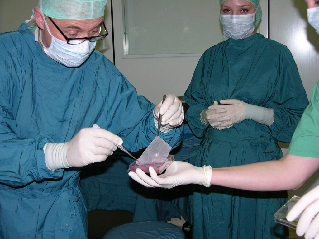
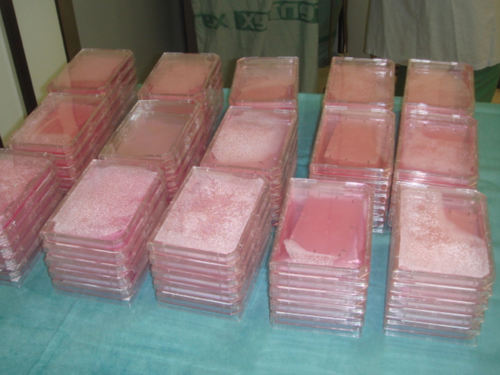
Since the 1980s, skin cells can be grown in the laboratory. The skin cells grown (cultured) in the laboratory can be inserted into the wound as cell layers or sprayed on the wound.
Split skin graft
For extensive burns and very superficial wounds, so-called split-thickness skin grafting is the treatment of choice. Split skin consists of an ultra-thin skin graft of the thickness of only 0.2 mm.
SkinDot skin graft
In the case of a deep wound with a loss of the entire skin, skin replacement with so-called full-thickness skin provides the best result. Full-thickness skin grafts are very stable due to their dermal shifting layer and contain all skin appendages.
Culture Skin Process and Skin Cell Cultivation
.
Cultured epidermal autografts (“CEA”) have been routinely used in the treatment of severely burn injured patients since 1981 [13]. However, it was the work of Cuono in 1987 and Hickerson in 1994, which additionally described allogeneic dermis in the wound bed prior to CEA transplantation, that improved the take rate of transplantation of laboratory cultured keratinocytes. Bilaminar skin substitute matrices from keratinocytes and fibroblasts are now commercially available. Research into tri- and multilaminar constructs of cultured cells for skin transplantation is being conducted worldwide, so that these tissue engineering procedures have now even developed their own terminology with the term “skingineering”. Further engineered dermal matrices and engineered dermo-epidermal matrices will become transplantable in the future. However, expectations for CEA have been clearly disappointed. Forty years after Rheinwald and Green, multiple dermal cell layers are not available because the skin as an organ with corresponding cell-to-cell communication has been completely underestimated.
Cultured Epithelial Autograft (CEA)
CEA grafts, which are now routinely used, consist of a multilayered sheet of the patient’s own keratinocytes (CEA sheets). The advantages are the almost unlimited number of cells that can be cultivated in a limited donor area of the severely burn injured patient. In the context of superficial wounds located in the dermis, these grafts lead to cosmetically flawless postoperative results. In contrast, when the dermis is lost and the graft is grafted to fat or muscle tissue, the results are extremely poor both functionally and aesthetically. It was not until work by Cuono in 1987 and Hickerson in 1994, who first transplanted allogeneic dermis into the wound bed, that better clinical results were achieved.
It is therefore important to establish dermal components in the wound bed before using CEA grafts in order to increase the take rate (healing rate) of cultured keratinocytes. In cases of full-thickness skin loss, this is achieved primarily by preparatory wound conditioning using glycerol- or cryopreserved allogeneic cadaveric or foreign skin. The foreign skin initially heals due to the immunosuppression associated with the severe burn injury and can serve as a substitute skin for the three- to four-week period of cell cultivation. In some cases, several surgical steps succeed in this way to fix allogeneic dermal components in the wound bed to allow transplantation of CEA after full-thickness skin loss.
This procedure can also be used to treat extensive burns of more than 90% body surface area.
| CEA Sheet | Vorteil | Disadvantage |
|---|---|---|
| Skin quality of the graft | Poor, because no skin appendages in the graft, no stem cells present, no dermal shift layer, consisting of keratinocytes only. | |
| Healing of the graft | Poor, as very sensitive to pressure | |
| Scarring | Gering | |
| Donor area | Small area removal for cell cultivation possible on all remaining healthy skin areas | |
| OP time | Complicated technique, long manufacturing time of the transplants in specially approved institutes necessary | |
| Costs | Very high cost, one sheet (10 x 10 cm = 400 Euro) |
Cultured Epithelial Autograft (CEA) als Suspension
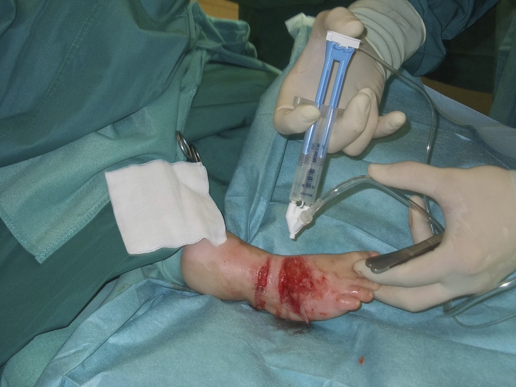
Transplantation is possible not only of differentiated, multilayered transplantable keratinocyte cell clusters as sheets, but also of the suspension of a predominantly poorly differentiated, proliferative keratinocyte population. Hunyadi et. al. were the first to suspend trypsinized, uncultured keratinocytes in fibrin glue on wounds [21,22]. Later, devices were added that allowed cell suspensions to be sprayed onto the wound to increase the uniform distribution of cells compared to transplantation by pipetting, resulting in the commercially available product Recell®. With this method, skin cells can be obtained directly after preparation from a no split skin piece and sprayed intraoperatively onto the wound as a mixed cell population without cultivation (spray application).
However, because of the dermal component required, the cell spraying method is only suitable for superficial second-degree wounds and has therefore not gained widespread acceptance in the treatment of severely burned patients.
The advantage of Recell® is rapid and safe wound closure in very superficial skin defects, especially superficial burns and scalds in the facial region. However, the transplanted mixed cell suspension is only successful in the presence of dermal components in the wound.
| Product name | Cultured Epithelial Autografts (CEA) | |
|---|---|---|
| Epicel® | Genzyme Biosurgery | Cultured autologous epidermal human keratinocytes, grown using mouse fibroblasts, Sheet. |
| Myskin® | Celltran Ltd | Cultured autologous epidermal human keratinocytes, cultured using irradiated mouse fibroblasts, sheet |
| Epidex® | Euroderm GmbH | Cultured autologous epidermal human keratinocytes, cultured using mouse hair follicle cells, sheet. |
| Keratinozytensheet | Deutsches Institut für Zell- und Gewebetransfer (DIZG) | Cultured autologous epidermal human keratinocytes, grown using collagen-coated cell culture flasks, Sheet. |
| Keratinozytensuspension | Deutsches Institut für Zell- und Gewebetransfer (DIZG) | Cultured autologous epidermal human keratinocyte suspension, cultured using collagen-coated cell culture flasks, suspension. |
| Recell® | Clinical Cell Culture Ltd | Autologous intraoperatively prepared autologous human mixed cell suspension. |
Do you want to offer the new, innovative SkinDot procedure to your patients? Contact us! We support you with joint surgery planning, surgery execution, matrices, and corresponding patient documents (informed consent form, risk exclusion). We look forward to hearing from you!
Skin Transplantation beyond
Skin Transplantation 2.0


